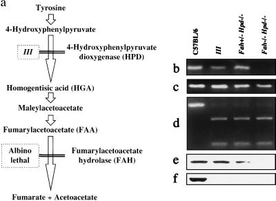
Figure 1.
Characterization of Fah−/− Hpd−/− double-mutant mice. (a) Scheme of tyrosine catabolism and the model mouse for tyrosinemia. The Fah−/−Hpd−/− mice were generated by intercrossing III and albino lethal mice. (b–d) Genotype analysis for Fah−/−Hpd−/− mice by PCR. Deletion in the c14cos mice disrupted the Fah gene, resulting in the absence of exon 1 and 2 sequences (7, 11, 14). The Fah−/− mice were identified by the absence of exon 2 sequences (111 bp) and the presence of exon 8 sequences (132 bp) of the Fah gene after PCR amplification of regions containing each exon, respectively (11). The results of PCR amplification of regions containing exon 2 (b) and exon 8 (c) of the Fah gene are shown. (d) Results of PCR amplification and restriction enzyme digestion of a region containing exon 7 of the Hpd gene. The left lane contains an undigested fragment with normal sequences (386 bp) after treatment with the restriction enzyme HindIII. Lanes 2–4 show products digested by HindIII because of the mutated sequences of the Hpd− allele (272 and 114 bp). DNA fragments were analyzed by electrophoresis on 4% NuSieve agarose gel (FMC BioProducts) and stained with ethidium bromide. (e and f) Immunoblot analysis of FAH (e) and HPD (f) protein in the liver. FAH and HPD proteins were absent from the liver from the Fah−/−Hpd−/− mouse.

