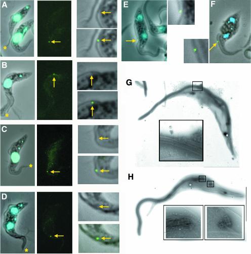
Fig. 5. The FC in wild-type and in induced (TbIFT88) RNAi cells. Normal wild-type (A–D) and (TbIFT88) RNAi trypanosomes induced for 36 h (E–F) were stained with an anti-FC antiserum. Left panels, phase contrast image merged with DAPI staining (blue). Central panels, FC staining only; right panels, magnification of the area surrounding the FC. Top, phase contrast image; bottom, phase contrast image merged with FC staining (green). Stars, tip of the old flagellum; arrows, tip of the new flagellum. (A) The left cell has only one flagellum, and its tip is not stained. The right cell has an elongating new flagellum and its distal tip shows defined staining. In contrast, the distal tip of the old flagellum is not stained. Similar observations were made as the new flagellum elongated (B–D) and stained with the anti-FC antiserum (green). (E) Binucleated cell whose new flagellum is too short (compare with D). (F) Binucleated cell without a new flagellum. FC is still present on the old flagellum of ∼30% of these cells. (G) Electron micrograph of a negatively stained cytoskeleton of (TbDHC1b) RNAi non-induced trypanosome. The discrete pyramidal structure of the FC is shown in the enlarged box. (H) Electron micrograph of a negatively stained cytoskeleton of (TbDHC1b) RNAi trypanosome induced for 48 h. The old basal body lies adjacent to the flagellum (enlarged left box), whereas no flagellum at all is visible on the new basal body (enlarged right box). In these conditions, the FC is not detected.

An official website of the United States government
Here's how you know
Official websites use .gov
A
.gov website belongs to an official
government organization in the United States.
Secure .gov websites use HTTPS
A lock (
) or https:// means you've safely
connected to the .gov website. Share sensitive
information only on official, secure websites.
