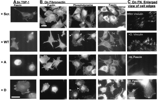
Fig. 8. Effects of TAT-FAS peptides on F-actin organization and matrix contact structures. C2C12 cells were preloaded for 1 h with 200 nM peptides as indicated, plated onto 50 nM TSP-1 (A), or 50 nM FN (B and C), for 1 h in the continued presence of peptides and fixed and stained for (A) fascin, or (B) F-actin, fascin or phosphotyrosine. Bars, 10 µm. (C) Higher magnification views of cell edges. Bar, 5 µm. Images are representative of six independent experiments.

An official website of the United States government
Here's how you know
Official websites use .gov
A
.gov website belongs to an official
government organization in the United States.
Secure .gov websites use HTTPS
A lock (
) or https:// means you've safely
connected to the .gov website. Share sensitive
information only on official, secure websites.
