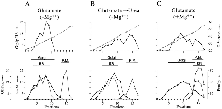
Figure 3.
In cells grown on glutamate, Gap1p-HA is in the ER and Golgi but not the plasma membrane. (A) Cell lysates from wild-type (CKY465) grown on glutamate, were fractionated on density gradients of 20–60% sucrose containing 10 mM EDTA. Relative levels of Gap1p-HA, Pma1p (plasma membrane marker), and Sec61p (ER marker) in each fraction were quantitated by Western blotting and densitometry. GDPase (Golgi marker) was determined by enzymatic assay of each fraction. The position of the peak fractions for each marker are indicated (these fractions contain at least 80% of the total marker on the gradient). (B) Cell lysates from wild-type (CKY465) grown on glutamate and then transferred to urea medium were fractionated on density gradients containing 10 mM EDTA. (C) Cell lysates from wild-type (CKY465) grown on glutamate were fractionated as above but on density gradients containing 2 mM Mg2+.

