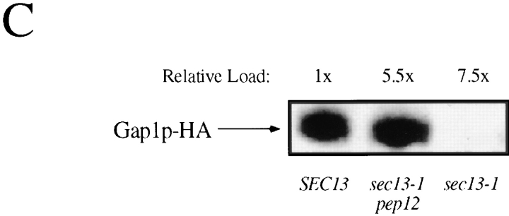
Figure 9.

Gap1p is transported from the Golgi to the vacuole in sec13-1 mutants. (A) Wild-type (CKY520), sec13-1 (CKY521), and sec13-1 pep4Δ (CKY467) strains were grown at 24°C to exponential phase. Cultures were labeled with [35S]methionine and [35S]cysteine for 10 min and chased by the addition of an excess of unlabeled methionine and cysteine. Gap1p-HA was immunoprecipitated from labeled extracts, resolved by SDS-PAGE, and was quantified using a phosphorimager. (B) Gap1p activity was assayed by the rate of [14C]citrulline uptake in wild-type (CKY443), sec13-1 (CKY444), sec13-1 pep12Δ::TRP1 (CKY455), sec13-1 end3-1 (CKY456), and sec13-1 pep4Δ::LEU2 (CKY457) strains. (C) Cell lysates from wild-type (CKY465), sec13-1 (CKY466), and sec13-1 pep12Δ::TRP1 (CKY468) strains were fractionated on density gradients of 20%–60% sucrose containing 10 mM EDTA. Fractions containing Pma1p were pooled, and Gap1p-HA in these fractions was detected by Western blotting with anti-HA antibody. The relative amount of plasma membrane in each extract was determined by quantitation of Pma1p. For A–C, all strains were grown in ammonia medium.

