
Figure 6.
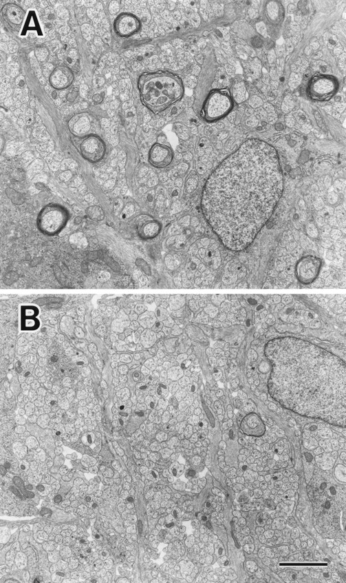
Electron micrographs showing representative areas of cross sections of optic nerves of MAP1B+/+ (A) and −/− (B) mice at postnatal day 8. Axons in the MAP1B−/− optic nerve (B) are less myelinated than those in the MAP1B+/+ optic nerve (A). Bar, 2 μm.