
Figure 8.
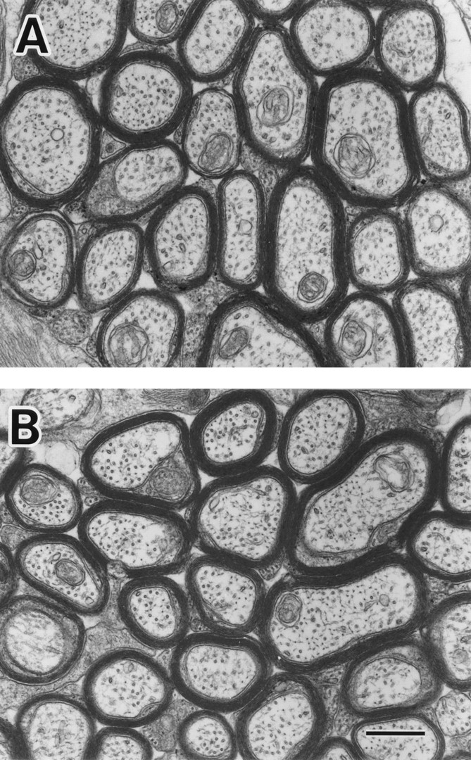
Electron micrographs showing representative areas of cross sections of optic nerves of MAP1B+/+ (A) and −/− (B) mice at postnatal week 14. There are no differences in the number of myelinated axons per unit area, diameter of axons, thickness of myelin sheaths, and density of MTs. Bar, 500 nm.