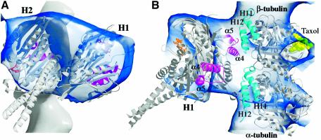
Fig. 4. Docking into a motor–microtubule cryoEM map. The new atomic structure of Ncd is shown docked into a cryoEM density map of nucleotide-free Ncd bound to microtubules (Hirose et al., 1998, 1999). (A) View perpendicular to the microtubule axis (similar view of motor as Figure 1B but rotated to the left). A microtubule protofilament is shown at the back with the plus end at the top (motor elements L11–α4–L12–α5, magenta; ADP, gold). (B) View from the side of the unattached head H1. An α/β tubulin atomic model (PDB accession code 1TUB) (Nogales et al., 1998) is also shown docked into the cryoEM map (helices H11 and H12, cyan; taxol, light green). Figure prepared with BOBSCRIPT (Esnouf, 1997).

An official website of the United States government
Here's how you know
Official websites use .gov
A
.gov website belongs to an official
government organization in the United States.
Secure .gov websites use HTTPS
A lock (
) or https:// means you've safely
connected to the .gov website. Share sensitive
information only on official, secure websites.
