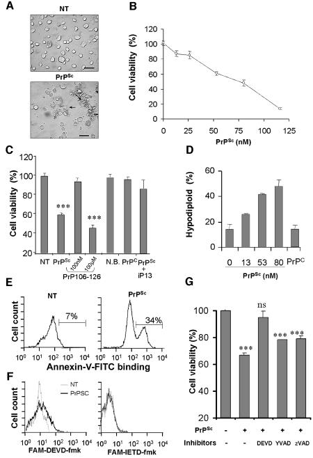
Fig. 1. Purified PrPSc from 139A-scrapie infected brains induces caspase-3 dependent apoptosis of N2A cells. (A) Morphological changes observed in N2A neuroblastoma cells treated with 50 nM PrPSc for 48 h. (B) N2A cells were treated with different concentrations of PrPSc and cell viability was analyzed after 48 h by MTS assay. (C) As controls, cells were incubated with: 165 nM of recombinant mouse PrPC; 50 nM PrPSc pre-treated with 300 nM of β-sheet breaker peptide iPrP13 to decrease PrPSc-β sheet content; 100 nM or 100 µM of aggregated PrP106–126 peptide for 7 days; or 0.5% (v/v) preparation of normal brain homogenate following the same procedure as to purify PrPSc (N.B.). (D) Cells were incubated for 48 h with different concentrations of PrPSc, 100 nM recombinant PrPC or left untreated. Subsequently, cells were stained with PI and hypodiploid cell population was quantified by FACS analysis. (E) Cells were treated with 66 nM PrPSc for 6 h and then phosphatidylserine exposure on the cell surface was detected using annexin V–FITC. (F) Caspase-3 and caspase-8 activity in situ was determined by flow cytometry using the cell permeable substrates FAM-DEVD-fmk and FAM-IETD-fmk after treatment with PrPSc for 20 h. Non-treated cells (gray line), or cells treated with 50 nM PrPSc (black line). (G) Cells were incubated for 60 min with or without 100 µM Ac-DEVD-fmk (DEVD), 100 µM Ac-YVAD-fmk (YVAD) or 10 µM zVAD-fmk (zVAD). Then, PrPSc was added to a final concentration of 50 nM, and after 48 h cell survival was determined by the MTS assay. Data shown in (C, D and G) correspond to the means and standard deviations at two independent experiments performed in triplicate. Statistical analysis was performed by parametric t-test comparing each value with the untreated control.

An official website of the United States government
Here's how you know
Official websites use .gov
A
.gov website belongs to an official
government organization in the United States.
Secure .gov websites use HTTPS
A lock (
) or https:// means you've safely
connected to the .gov website. Share sensitive
information only on official, secure websites.
