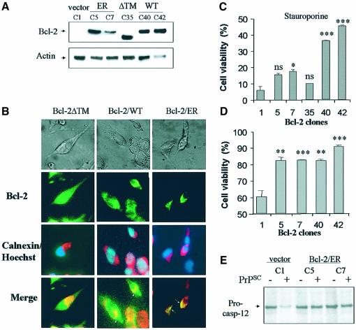
Fig. 4. Bcl-2 targeted to the ER decreases the sensitivity of N2A cells to PrPSc toxicity and caspase-12 activation. (A) Cells were stably transfected with Bcl-2/ER, Bcl-2-ΔTM and Bcl-2WT constructs or with empty vector, and Bcl-2 protein levels were analyzed by western blotting in different cellular clones. (B) In parallel, Bcl-2 distribution was detected by immunofluorescence in selected clones. Bcl-2 staining is shown in green; calnexin staining is shown in red and nuclear labeling with Hoechst33342 is shown in blue. (C) As controls, Bcl-2 transfected clones and control cells were treated with 150 nM staurosporine for 24 h. (D) The same N2A cell clones were treated with 50 nM PrPSc for 48 h, and cell viability was assessed using MTS analysis. In the last two panels, statistical analysis was performed comparing the cell viability values for each transfected clone with the results obtained with the control clone C1. (E) Cells transfected with vector alone (C1 clone) or with Bcl-2/ER (clones 5 and 7) were treated with 50 nM PrPSc. Caspase-12 activation was analyzed by western blot. The significance of the differences in pro-caspase-12 expression was estimated by densitometric analysis of the blots.

An official website of the United States government
Here's how you know
Official websites use .gov
A
.gov website belongs to an official
government organization in the United States.
Secure .gov websites use HTTPS
A lock (
) or https:// means you've safely
connected to the .gov website. Share sensitive
information only on official, secure websites.
