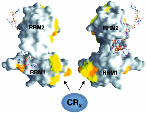
Fig. 8. Molecular surface representation of dSXL RRMs bound to RNA. Two different orientations, vertically rotated by 180°, are shown (Handa et al., 1999). Residues that differ between Drosophila and Musca SXL are highlighted in yellow (conservative changes) and orange (non-conservative changes). A bound RNA oligomer derived from the tra mRNA is depicted using a stick representation. The solvent-exposed surface of RRM1, containing a high proportion of residues that differ from Musca SXL, is suggested to interact with translational co-repressors.

An official website of the United States government
Here's how you know
Official websites use .gov
A
.gov website belongs to an official
government organization in the United States.
Secure .gov websites use HTTPS
A lock (
) or https:// means you've safely
connected to the .gov website. Share sensitive
information only on official, secure websites.
