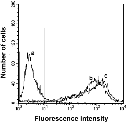
Figure 4. The binding of FITC-labeled TP5 onto the surface of EBV-transformed B cells.
EBV-transformed B cells were incubated with PBS alone (trace a), FITC-labeled TP5 (trace b) and mAb-DR FITC (trace c), and then subjected to flow cytometry. The vertical line was the boundary between bound and unbound cells.

