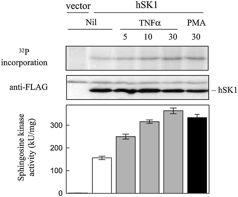
Fig. 1. Agonist-induced phosphorylation of hSK1. Time course of in vivo hSK1 phosphorylation and activity following TNFα (1 ng/ml) and PMA (10 ng/ml) stimulation of cells. HEK293T cells over expressing hSK1 were metabolically labelled with 32P prior to treatment with TNFα and PMA for the indicated times (min). Immunoprecipitated hSK1 from these cells was then subjected to SDS–PAGE and the incorporation of 32P into hSK1 determined. Loading controls for hSK1 in the immunoprecipitates were visualized by western blot via their FLAG epitope. Quantitation of 32P incorporation into hSK1 following 5, 10 and 30 min of TNFα treatments showed fold increases of 1.7 ± 0.3, 2.3 ± 0.3 and 3.4 ± 0.4, respectively, over that seen in the untreated cell extracts. Similarly, PMA treatment for 30 min resulted in a 2.9 ± 0.3-fold increase in 32P incorporation into hSK1 compared with the untreated cell extracts. Sphingosine kinase activities in the extracts were determined prior to immunoprecipitation. All data are represented as means (± SD) from more than three experiments.
