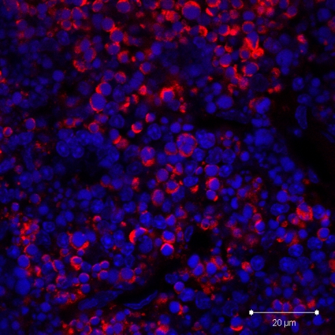
Fig. 3.
Paraffin section of thymus from mouse treated with dexamethasone and stained with anti-cleaved caspase 3 antibody (red). Confocal microscopy image shows intensely stained cells in the cortex, with neighboring unstained cells. Nuclei have been stained with DAPI (blue). This represents a good positive control for the anti-cleaved caspase 3 antibody technique

