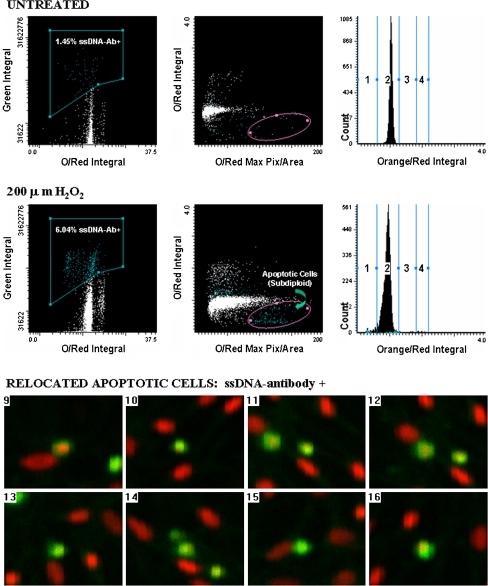
Fig. 4.
Laser scanning cytometry determination of apoptosis in lung epithelial cells using anti-ssDNA antibody. Upper panel scattergrams showing percentage of ssDNA-positive cells (green integral vs. orange/red integral), the apoptotic fraction (orange/red integral vs. orange/red maximum pixel), and a DNA histogram for untreated C10 cells. Middle panel scattergrams showing percentage of ssDNA-positive cells (green integral vs. orange/red integral), the apoptotic fraction (orange/red integral vs. orange/red maximum pixel), and a DNA histogram for C10 cells treated with 200 μM H2O2 for 24 h. Note the increased number of cells ssDNA-positive, and the enhanced subdiploid DNA fraction in cells treated with H2O2 as compared with sham controls. Bottom panel cell relocation feature of LSC demonstrated for eight cells. Cells within the subdiploid fraction (elliptical region highlighted on scattergram in middle panel) were relocated and visually confirmed as apoptotic by morphological appearance and positive staining with the ssDNA antibody. Reprinted with permission from BioTechniques (Taatjes et al. 2001)

