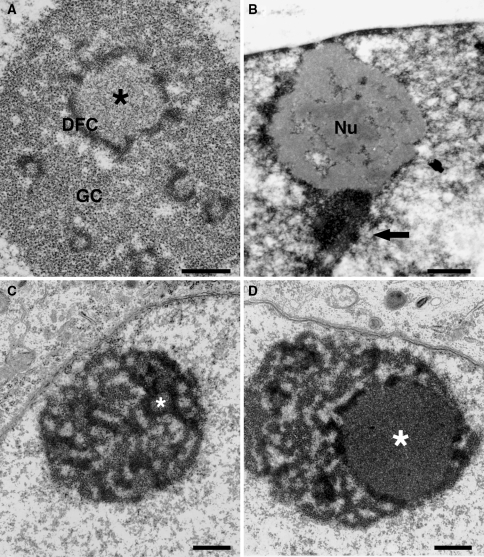
Fig. 1.
The nucleolus of mammalian cells as seen by electron microscopy. a In the human HeLa cell, the three main nucleolar components are visible in a section of material fixed in glutaraldehyde and osmium tetroxyde, embedded in Epon and the section contrasted with uranyl acetate and lead citrate. FCs of different sizes are visible and the largest is indicated by an asterisk. The FCs are surrounded by the DFC and are embedded in the GC. b Preferential contrast of DNA using NAMA-Ur staining in a PtK1 cell (courtesy J. Gébrane-Younès). The nucleolus is the gray structure surrounded by highly contrasted chromatin (arrow). Some chromatin filaments are also visible inside of the nucleolus (Nu). c, d Nucleolus of rat neurones (courtesy M. J. Pébusque) in the day (c), and during the night (d) which is the active period for the nucleolus of the rat. In the nonactive period (c), the nucleolus is reticulated with small FCs (asterisk). In the active period, one giant FC is visible (d, asterisk). Bar in a: 0.5 μm and bars in b, c and d: 1 μm

