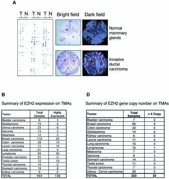
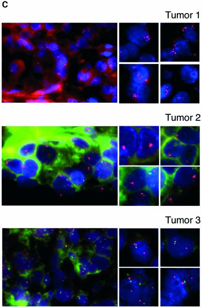
Fig. 5. EZH2 is highly expressed and amplified in primary human tumors. (A) EZH2 is highly expressed in primary breast tumors. The expression of EZH2 mRNA levels was determined by ISH on TMA. The left panel shows a representative example of ISH for EZH2 of a breast cancer TMA. Two samples from each tumor (T) and normal (N) counterparts were taken for the TMA. The bright field panels show the morphology of the tissue sample as revealed by hematoxylin and eosin counterstaining. The dark field panels show the ISH analysis of EZH2 expression levels in a breast invasive ductal carcinoma and in a normal mammary gland. In the panels the numbers indicate: 1, stroma; 2, mammary gland epithelial cells; 3, tumor tissue; 4, EZH2 expressing cells. (B) EZH2 is highly expressed in a large number of primary human tumors. Summary table of EZH2 expression on the TMAs tested. (C) Subsets of primary tumors show an amplification of the EZH2 gene (top and middle panel). FISH analysis of tumor TMAs of EZH2 gene copy number. The two panels show two representative breast carcinoma tissues with significant amplifications of EZH2. The EZH2 specific probe was labeled with Cy5 and a centromeric probe for chromosome 7 with FITC. Due to the fast fading of the FITC probe and the relative long microscopic analysis of each TMA we were able to count, but not to take pictures of the specific staining of the centromere for several of the analyzed samples. (C, lower panel) Example of a breast tumor tissue containing a normal copy number of EZH2. FISH analysis of an unrelated gene (HECTH9) on the same TMAs revealed no amplification in all the samples tested (data not shown). (D) Summary table of EZH2 copy number of the tested TMAs.

An official website of the United States government
Here's how you know
Official websites use .gov
A
.gov website belongs to an official
government organization in the United States.
Secure .gov websites use HTTPS
A lock (
) or https:// means you've safely
connected to the .gov website. Share sensitive
information only on official, secure websites.

