
Figure 1.

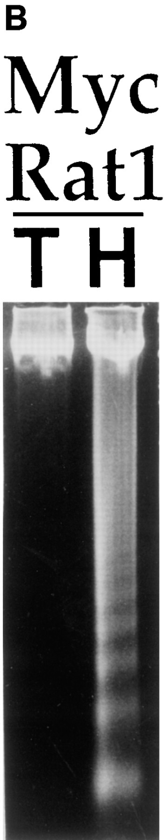


Rat embryo fibroblasts transformed with Myc/Ras or E1A/Ras are triggered into apoptosis when deprived of substrate adhesion. (A) Oligonucleosomal genomic DNA fragmentation. Early passage primary REF and Myc/Ras-REF were plated on either tissue culture plastic (lanes 2 and 5), bacterial petri (3 and 6), or polyHEMA-coated TCP dishes (4 and 7), and genomic DNA was isolated from 5 × 10⁶ cells as described in the Materials and Methods. (B) Myc/Rat1 cells were plated on TCP (T) or polyHEMA (H) as above, and genomic DNA was isolated from 5 × 10⁶ cells as described in Materials and Methods. (C) E1A/Ras-REF and parental REF were plated on TCP or polyHEMA-coated TCP for 12 h before nuclear staining with DAPI as described in Materials and Methods. (D) REF clones transformed with oncogenes Myc/Ras or E1A/Ras or early passage primary REF were plated at 105 cells/100-mm dish on TCP (open bars) or polyHEMA-coated TCP (closed bars). Viability was measured 12 h after plating by calculating the ratio of trypan blue-positive cells to live cells (see Materials and Methods).