
Figure 1.

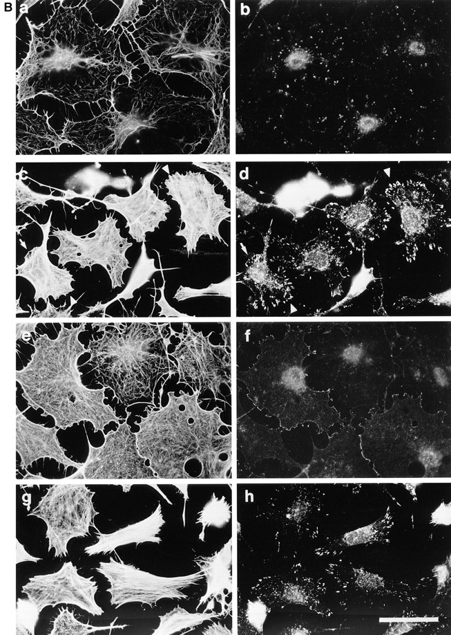
Permeabilization of quiescent Swiss 3T3 cells in the presence of GTPγS. (A) Permeabilization (protocol 1) was performed in the absence of stimulus (left) or in the presence of 50 μM GTPγS (right). Cellular F-actin was visualized using rhodamine-conjugated phalloidin. (B) Permeabilization (protocol 1) was performed in the absence of stimulus (a and b), in the presence of 50 μM GTPγS (c and d); 50 μM GTPγS with 0.1 nM C3 transferase (e and f); 50 μM GTPγS with 1 nM N17Rac (g and h). After 20 min at 37°C, cells were fixed and F-actin visualized with rhodamine-phalloidin (a, c, e, and g) and vinculin visualized with a monoclonal antibody (b, d, f, and h). In c and d, arrowheads show termini of bundled actin filaments decorated with focal adhesions, while arrows mark regions of peripheral actin polymerization decorated with linear arrays of focal complexes. Bars: (A) 150 μm; (B) 30 μm.