
Figure 3.
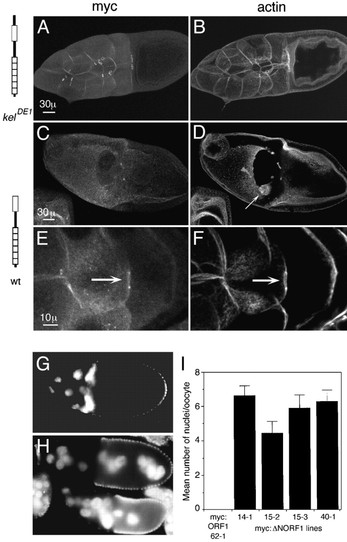
Dominant-negative effect caused by myc:ΔNORF1. (A) Myc:ORF1 localizes normally to ring canals. Some protein is also observed localizing to the cortical cytoskeleton. (B) The ring canals and nurse cell plasma membranes in kelDE1 egg chambers that express myc:ORF1 are intact and normal, as seen by visualizing filamentous actin. (C–F) Wild-type egg chambers expressing myc:ΔNORF1. (C) In wild-type stage 10 egg chambers that express myc:ΔNORF1, the nurse cell plasma membranes are severely disrupted and in this case missing. No ring canals are observed by staining for myc:ΔNORF1. (D) Actin staining of the same egg chamber as in C verified that the nurse cell membranes are completely disrupted. The border cells (arrow) have migrated toward the oocyte but are mislocalized, probably because there is no place for them to dock. (E) An occasional ring canal persists in this stage 9 egg chamber, and myc:ΔNORF1 (arrow) can be seen localizing to it. (F) Actin is also localized to the same thin ring canal. Cortical actin is also seen on some of the nurse cell membranes. (A, C, and E) Anti-myc staining to detect the transgene product. (B, D, and F) Rhodamine-conjugated phalloidin staining to detect filamentous actin. (G) A wild-type egg chamber expressing myc:ORF1 has normal positioning of the nuclei in the nurse cells. (H) Wild-type egg chambers expressing myc: ΔNORF1 have nuclei transported into the oocyte. (G and H) Nuclei are detected by DAPI staining. (I) Comparison of the number of nuclei transported into the oocytes in four independent wild-type lines that express myc:ΔNORF1. No nuclei were observed transported into the oocytes in wild-type egg chambers that express myc:ORF1 (line 62-1). 17–30 stage 10 or older egg chambers from 5–10 females of each line were counted for the averages. Histograms represent the means, and the error bars represent the standard error of the mean. The phenotype was observed in several additional trials although they were not quantitated. Bars: (A and C) 30 μm; (E) 10 μm.