
Figure 4.
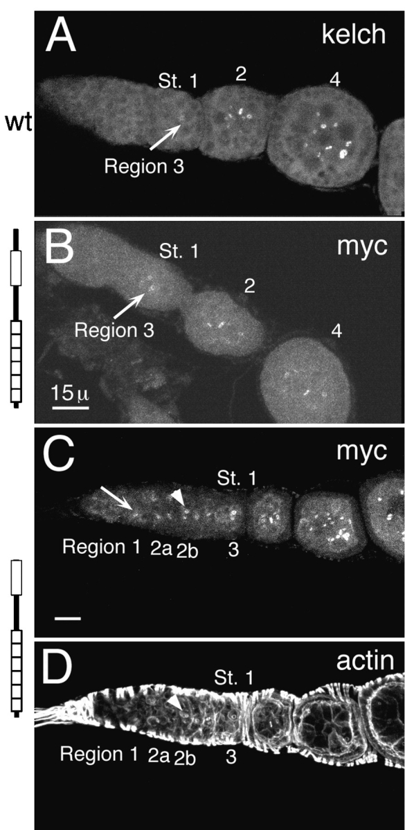
Myc:ΔNORF1 localizes to ring canals earlier than normal. (A) Endogenous kelch localized to ring canals (arrow) beginning in region 3/stage 1. It takes until stage 4 for it to be detected on all of the ring canals in the egg chamber. This ovariole was stained with anti-kelch 1B antibodies. (B) Myc:ORF1 began to localize to ring canals (arrow) also in region 3/stage 1 egg chambers. It also took a few stages to reach all of the ring canals in the egg chamber. (C) Myc:ΔNORF1 began to localize to region 1 ring canals (arrow). The ring canals identified by the arrowhead correspond to those identified by the arrowhead in D. B and C were stained with anti-myc 9E10 antibodies to recognize the transgene products. (D) The same germarium as in C stained with rhodamine-conjugated phalloidin reveals the accumulation of the robust inner rim of actin filaments. The inner rim of actin filaments begin to accumulate by region 2a but are not really obvious until region 2b (arrowhead). The germaria in B–D are kelDE1 mutants expressing the respective transgene. Bars, 15 μm.