
Figure 11.
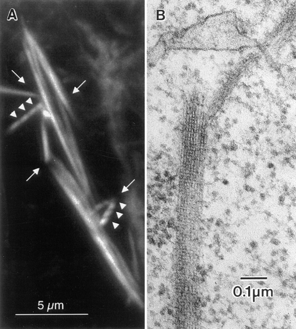
(A) Portion of a kelchneo stage 10 egg chamber stained with rhodamine-conjugated phalloidin and examined by confocal microscopy. A number of striated actin bundles can be seen (arrowheads). Of interest here are the distorted regions characterized by angular intersections of relatively straight bundles (arrows). Similar distortions can be seen in wild-type egg chambers, especially where the actin bundles contact the nuclear envelope (e.g., Fig. 2 b). This is a 3-μm optical section. (B) Thin-section through the portion of the nurse cell cytoplasm from a kelchneo stage 11 egg follicle. Of interest is that the cable appears broken. This occurs at the point of overlap of two modules. Bars: (A) 5 μm; (B) 0.1 μm.