
Figure 5.
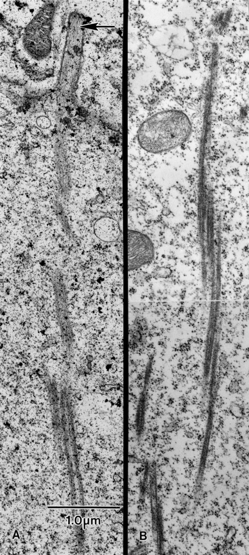
Thin-sections through a portion of the surface of a wild-type nurse cell. (A) Extending basally from a microvillus (arrow) is an actin filament bundle. This bundle, as it extends basally, is connected to other actin bundles that run parallel to it. (B) Thin-section cut deeper in the cytoplasm. This micrograph illustrates that actin bundles associate laterally with other bundles. Although it cannot be proven from these two micrographs, individual bundles appear short (e.g., 2–3 μm) but overlap like the units of an extension ladder. Both A and B are cut from stage 11 follicles and printed at the same magnification. Bar, 1 μm.