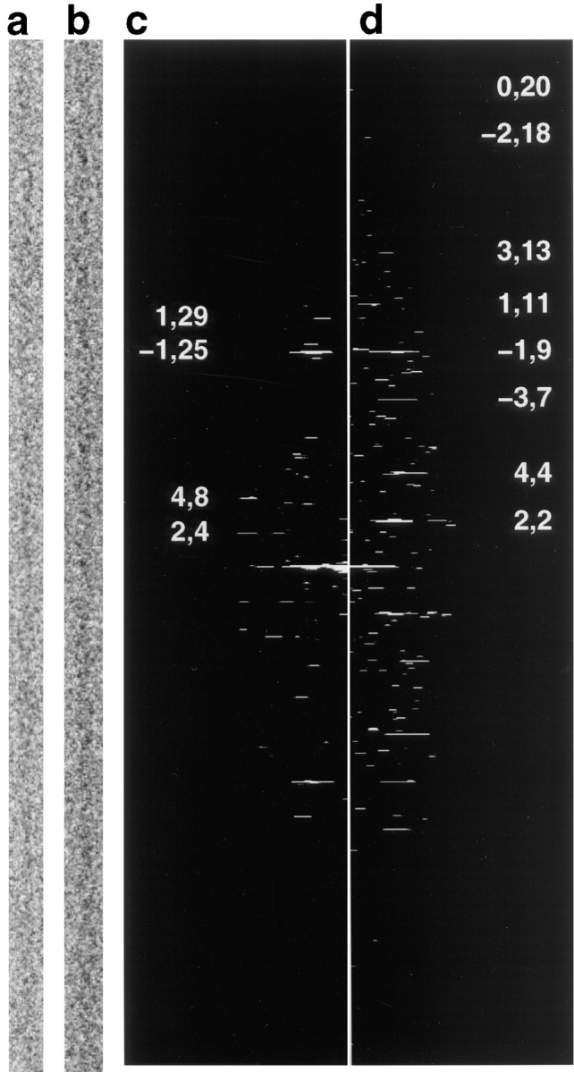
Figure 2.

Cofilin changes the twist of platelet F-actin. (a) Portion of a computationally straightened F-actin filament; (b) portion of a computationally straightened actin filament decorated with cofilin. The defocus of both micrographs shown was 1.5 μm. Decorated filaments are ∼120–130 Å diam with accentuated ‘crossovers' relative to control actin. (c) Computed diffraction pattern calculated from the actin filament shown in (a). The length of the filament used to calculate this pattern was 1.44 μm. (d) Computed diffraction pattern calculated from the cofilin-decorated actin filament shown in (b). The length of the filament used to calculate this pattern was 1.41 μm. Prominent layerlines in each diffraction pattern are labeled with values of n and l. The axial position of layerline n = 2, l = 4 in the F-actin diffraction pattern is 1/366 Å−1 and that of n = 2, l = 2 in the cofilin/F-actin diffraction pattern is 1/272 Å−1.
