
Figure 9.
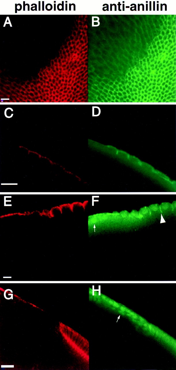
Defects arise at the onset of cellularization and persist throughout in embryos from syx L266 clones. As soon as membrane invagination begins in mitotic cycle 14, defects in the actin cytoskeleton are apparent (A and C). Anillin still shows a hexagonal grid early in cellularization (B), although it does not appear enriched in areas lacking F actin (D). The acellular patch begins near the left edge of C and D. By the midpoint of cellularization, defective regions still show no actin organization (E). Anillin is also disorganized in these regions, and shows a stronger nuclear localization than usual (small arrow, F). Anillin still localizes to the invaginating furrow in those regions undergoing cellularization (large arrowhead F). By the very late stages of cellularization, cytoskeletal organization is still completely lacking in defective regions (G). Anillin is almost completely localized to the nuclei in these regions (arrow, H). Bars: (A and B, C and D, and E and F) 10 μm; (G and H) 20 μm.