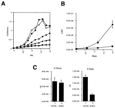
Figure 4.
Growth of H37Rv Δacr∷hpt in vitro and in vivo. (A) Growth of the parent (open symbols) and H37Rv Δacr∷hpt (closed symbols) under defined oxygen conditions. Each pair of lines represents growth under a continuous flow of a premixed oxygen in nitrogen atmosphere at 20% (squares), 5% (circles), 2.5% (triangles), and 1.25% (diamonds). Growth was monitored daily by optical density at 650 nm. (B) Growth of wild-type H37Rv (squares) and H37Rv Δacr∷hpt (circles) in THP-1 cells. Each point is a mean of total bacilli per well in triplicate wells, and the result shown is typical of several repetitions of this experiment. (C) Growth of the wild-type H37Rv (bars 1) and H37Rv Δacr∷hpt (bars 2) in primary mouse bone-marrow derived macrophages. (Left) Colony-forming units are shown immediately following infection (2 hr after infection) and on the Right 6 days later. The results shown are the average of triplicate wells plated at multiple dilutions.

