
Figure 6.
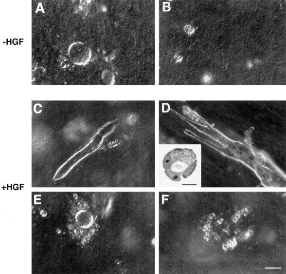
Tubulogenesis by LLC-PK1 cell transfectants within collagen gels. Spherical cysts were formed by control LLC-PK1 cells (A) and LLC-PK1 cells overproducing wild-type ezrin, grown under control conditions for 3 d. Small colonies were formed by LLC-PK1 cells overproducing the double phosphotyrosine-mutated ezrin-FF1 cells (B), and by cells overproducing the NH2-terminal domain of ezrin under the same growth conditions. In the presence of 30 ng/ml of HGF, some cysts developed into tubules in LLC-PK1 cells (C); the cysts developed in elongated tubules (three- to fivefold the control LLC-PK1 cells) in E7 cells (D). Inset represents a section of tubules formed and demonstrates lumen formation. N2 cells (E) or the tyrosine mutants (F) did not form tubules. Bars: (A–F) 100 μm; (inset) 25 μm.