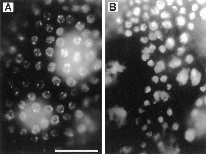
Figure 6.
Follicle cell defects in ovaries from KLP38B mutant females. Wild-type (Oregon R) stage 5–7 egg chambers have an even layer of follicle cells, revealed by DAPI staining (A). Comparable egg chambers from KLP38B 24-O females (B) have less follicle cells, leading to an uneven distribution of the follicle cells and large gaps in the layer. Exposed nurse cells with no overlying follicle cells are clearly visible. The follicle cell nuclei are of uneven size. This may be a consequence of earlier mitotic defects, but it may alternatively indicate uneven polyploidization of these nuclei. Nurse cells are the much larger nuclei beneath the follicle cell layer. Bar, 20 μm.

