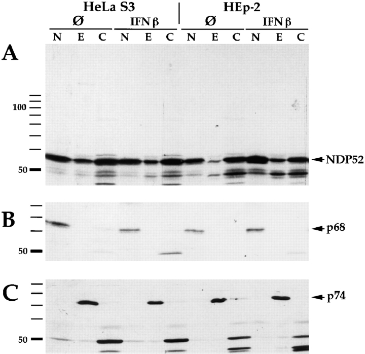
Figure 5.
Nucleo-cytoplasmic fractionation of HEp-2 and HeLa S3 cells. Cell fractions were separated by SDS-PAGE and analyzed by immunoblotting: Lanes labeled N, nuclear proteins; lanes labeled E, nucleus/cytoskeleton-associated proteins (designated E for extraction); lanes labeled C, the soluble cytoplasmic fraction. A shows detection of NDP52 (indicated by arrow) using rabbit anti-NDP52 antiserum. NDP52 is detected in all three subfractions. No redistribution is observed after treatment of the cells with IFN β. As a control for purity of the subcellular fractions, immunoblotting of the same extracts was performed using human autoimmune sera recognizing the snRNP p68 protein, which localizes in the nucleus (B, arrow) or the mitochondrial p74 protein, which localizes in the cytoskeleton fraction (C, arrow).

