
Figure 4.
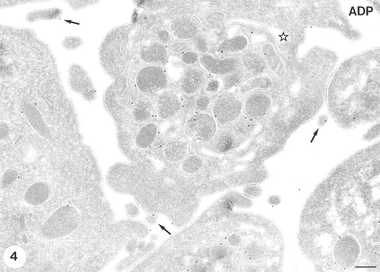
GLUT-3 expression after stimulation with 10 μM ADP. The shape changes characteristic for ADP stimulation are observed (i.e., centralization of α-granules and some cell surface ruffling ). A similar morphology is observed when the low dose of 0.05 U/ml thrombin is used (not shown). The majority of the label is still associated with the membranes of the α-granules. Some label is observed at developing pseudopods. Bar, 200 nm.