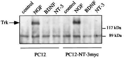
Figure 2.
Western blot analysis of neurotrophin-induced Trk activation. PC12 cells or PC12 cells stably transfected with pBJ-5-NT-3 myc were incubated with DMEM/0.1% BSA (control) or DMEM/0.1% BSA containing NGF, BDNF, or NT-3 (100 ng/ml each) for 5 min, lysed, and subjected to immunoprecipitation by using a polyclonal anti-panTrk antiserum. After SDS/PAGE and transfer to nitrocellulose membranes, Western analysis was performed by using antiphosphotyrosine antibodies. The arrow indicates tyrosine phosphorylated TrkA.

