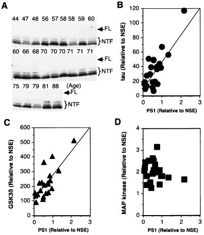
Figure 1.
Correlation between levels of PS1, tau, GSK-3β, and MAPK in human brains of various ages. The Triton X-100 soluble fraction of brain homogenate (60 μg total protein/lane) was examined by Western analysis using anti-PS1 antibody (MKAD3.4) (1:5), anti-tau antibody (JM) (1:5,000), anti-GSK-3β (Transduction Laboratories, Lexington, KY) (1:1,000), anti-MAPK (Santa Cruz) (1:2,000), and anti-NSE (Dako) (1:500). (A) Western blot using the mAb MKAD3.4. Top of each lane shows age of each person. MKAD3.4 reacts with a faint 47 kDa full-length PS1 (FL) and 28–35 kDa N-terminal fragments (NTF). The reactive band was quantified with a Densitograph Lumino-CCD (ATTO Corporation, Tokyo, Japan). The levels of PS1, tau, GSK-3β, and MAPK were normalized to the level of NSE in each sample, and the levels of tau (B), GSK-3β (C), and MAPK(D) were plotted against the corresponding PS1 levels. There were significant correlations between both PS1 and tau (n = 23, slope of regression line = 41.8, R2 = 0.65, P < 0.0001), as well as GSK-3β (n = 23, slope of regression line = 110.9, R2 = 0.61, P < 0.0001). In contrast, a linear regression showed no significant correlation between PS1 and MAPK (n = 23, slope of regression line = −0.2, R2 = 0.04, P > 0.1).

