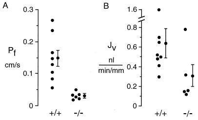
Figure 1.
Pf and Jv in isolated microperfused proximal tubules. S2 segments of proximal tubule were dissected from wild-type [+/+] and AQP1 knockout [−/−] mice and microperfused in vitro at 37°C as described in Methods. (A) Osmotic water permeability. Tubules were perfused with 295 mosM buffer and bathed in a 345 mosM buffer to give a 50 mosM bath-to-lumen osmotic gradient. Pf was computed from the increase in concentration of a membrane-impermeant marker at the distal end of the tubule. (B) Near-isosmolar fluid reabsorption. Tubules were perfused and bathed in an isosmolar buffer. Jv was computed from the increase in luminal marker concentration. For A and B, each point is the averaged data from 1 or 2 tubules from one mouse. Averaged data with SEs (n = number of different mice) is shown at the right of each data set.

