
Figure 1.


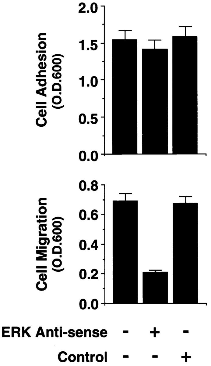

MAP kinase (ERK1 and ERK2) is required for cell migration but not adhesion or spreading on extracellular matrix proteins. (A, lower panel) Cells were allowed to adhere for 1 h or migrate for 6 h on a collagen substrate after exposure to MAP kinase–specific antisense or a scrambled control oligonucleotide. Migration studies were performed using Transwell migration chambers coated with collagen as described in Materials and Methods. (Upper panel) Total cell lysates (50 μg) prepared from an aliquot of those FG cells analyzed for migration/adhesion were immunoblotted with antibodies to Raf-1, actin, or ERK proteins. The upper band is a nonspecific protein recognized by the rabbit polyclonal ERK antisera. The result shown is a representative experiment from at least three independent experiments. (B, upper panel) Phosphotyrosine immunoblot or kinase activity of ERK1 and ERK2 proteins immunoprecipitated from cells either pretreated in the presence or absence of the MEK inhibitor PD98059 (25 μM) and allowed to attach to poly-l-lysine or collagen-coated dishes for 30 min. The ERK immunoprecipitates were also examined for their ability to phosphorylate MBP in an in vitro kinase assay as described in Materials and Methods. (Lower panel) Cells treated in the presence or absence of the MEK inhibitor as above were allowed to attach for 1 h or migrate for 6 h on a collagen substrate and quantified as described in Materials and Methods. The result shown is a representative experiment from at least three independent experiments. (C) Nomarski photomicrograph (600×) of FG cells pretreated for 6 h in the presence or absence (control) of the MEK inhibitor (100 μM) and allowed to attach and spread on collagen-coated coverslips for 10 or 60 min. The result shown is a representative experiment from at least three independent experiments. Bar, 10 μm.