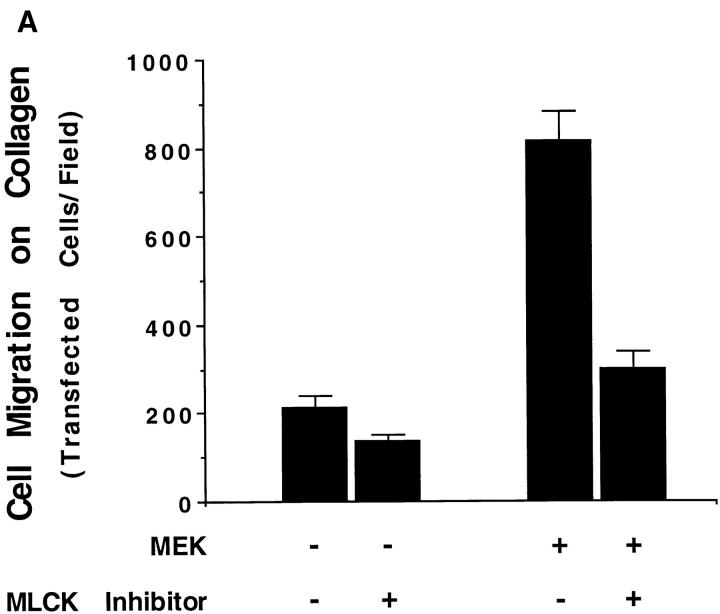
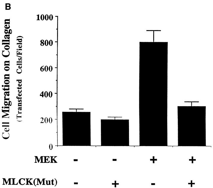
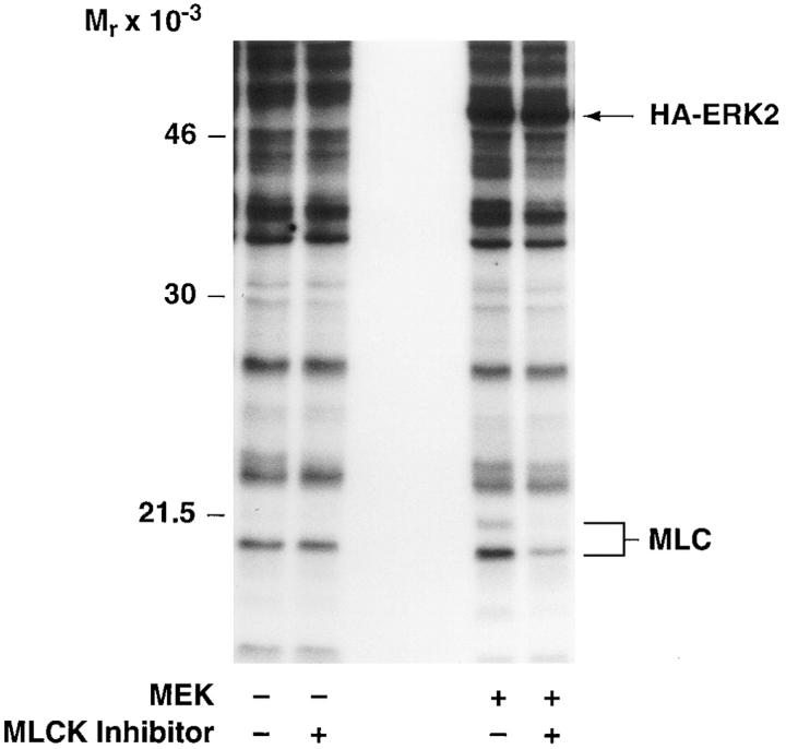
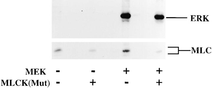
Figure 7.
MLCK activity is required for MAP kinase-induced phosphorylation of MLC and cell migration. (A, upper panel) COS cells were transfected with empty vector or mutationally active MEK+ along with an HA-tagged ERK2 reporter construct. Cells were allowed to migrate on a collagen substrate for 3 h in the presence or absence of the MLCK inhibitor KT5926 (10 μM) as described in Materials and Methods. Cell migration was enumerated as described above. (Lower panel) Lysates prepared from these COS cells metabolically labeled with 32P were examined by SDS-PAGE and autoradiography. The position of HA-tagged ERK and MLC are denoted by arrows. (B, upper panel) COS cells were transfected with mutationally active MEK+ and/or MLCK(Mut) and allowed to migrate on a collagen substrate as described in Materials and Methods. Cell migration was quantified by counting the number of migrant cells per high powered microscopic field as described above. (Lower panel) Lysates prepared from 32P metabolically labeled COS cells transfected as above were examined for phosphorylation of HA-ERK and MLC. The result shown is a representative experiment from at least three independent experiments.


