Figure 4.

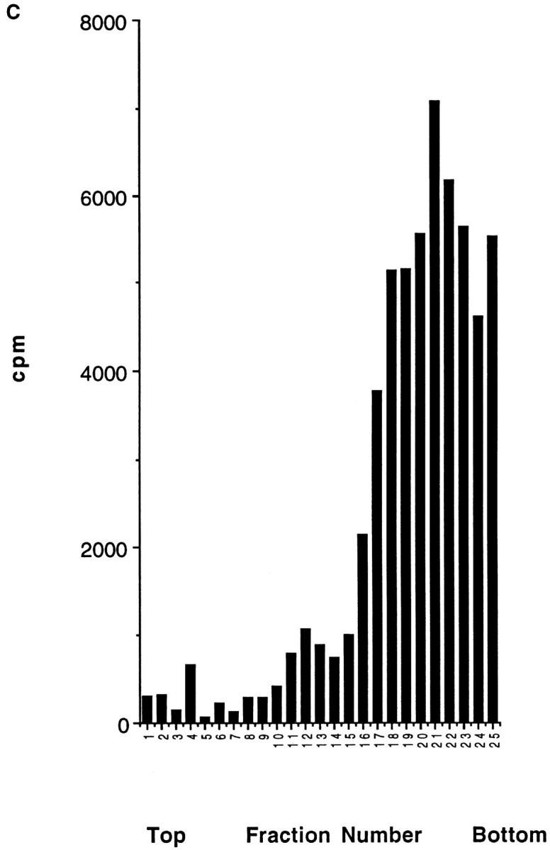
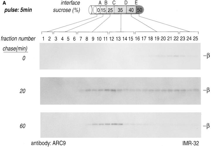
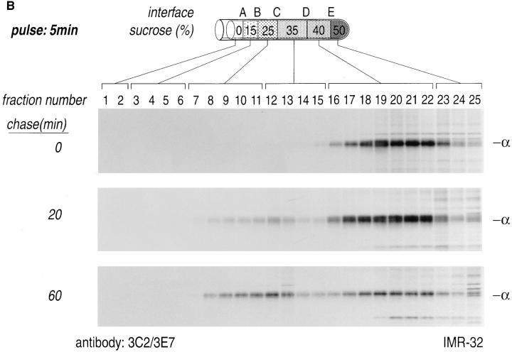
Domain localization of newly synthesized Gα and Gβ subunits. IMR-32 cells were pulse-labeled for 5 min with 500 μCi [35S]methionine and chased for the designated times. Cells were homogenized as shown (Fig. 3) and resolved on a discontinuous sucrose gradient. 0.5-ml fractions were collected from the top of each gradient, lysed in an equal volume of 2× NP-40/Lubrol lysis buffer, and aliquots from each fraction were immunoprecipitated with ARC9 (+ 0.2% SDS) (A) or with a mixture of anti-Gα antibodies (3E7, 3C2) (B). The interfaces between different sucrose densities are designated A–E, whereas the sucrose density steps are represented in percent sucrose. The distribution of radioactivity along the gradients is shown in C. Data shown are representative for at least three experiments performed in duplicate.


