
Figure 2.

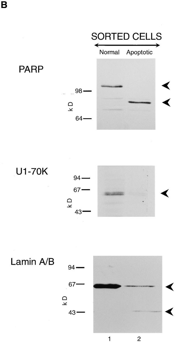
CPP32 and Ich-1 are extensively cleaved in apoptotic but not morphologically normal THP.1 cells, and their processing is concomitant with the cleavage of PARP, U1-70K, and lamins A/ B. THP.1 cells were incubated for 4 h in the presence of etoposide (25 μM), stained with Hoechst 33342 and propidium iodide, and then sorted by flow cytometry as described previously (Zhu et al., 1995). Cells with low blue fluorescence were morphologically normal and those with high blue fluorescence exhibited distinctive apoptotic morphology when examined by fluorescence microscopy. Cells with either normal (lane 1) or apoptotic (lane 2) morphology were analyzed by Western blot analysis as described in Materials and Methods. (A) Cells were analyzed using antibodies to CPP32 (upper panel) and Ich-1 (lower panel). (B) Cells were analyzed using antibodies to PARP, U1-70K, and lamins A/B (upper, middle and lower panels, respectively). The proforms of CPP32 and Ich-1 are indicated by the upper arrowheads (A), and intact PARP, U170K, and lamins A/B are indicated by the upper arrowheads (B). The lower arrowheads represent either processed enzymes or cleaved proteolytic fragments. Cells displaying normal morphology contain primarily the intact forms of all the proteins analyzed (lane 1), whereas, in apoptotic cells, processing of more than one ICE-like protease is detected and associated with cleavage of PARP, U1-70K, and lamins A/B.