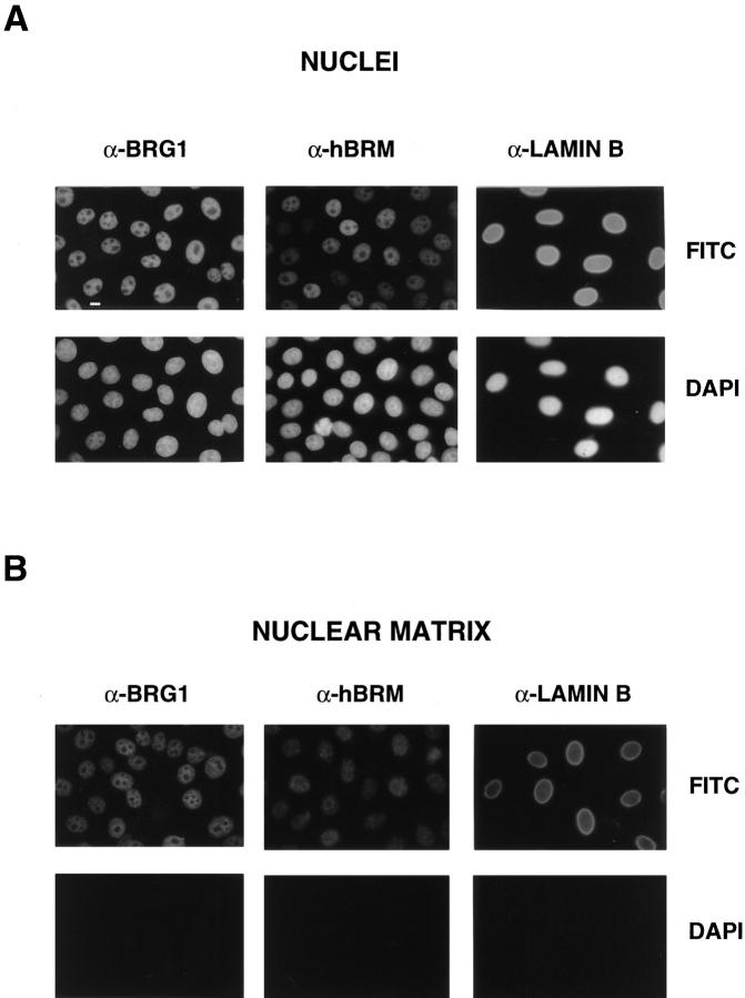
Figure 3.
In situ extraction of nuclear matrix. (A) Immunofluorescent staining of nonextracted HeLa cells with α-BRG1, α-hBRM, and α-lamin B antibodies. (B) Immunofluorescent staining pattern of in situ prepared nuclear matrices with α-BRG1, α-hBRM, and α-lamin B antibodies. Microscopy and photography parameters were standardized for all of the images. Bar, 10 μm.

