Figure 5.
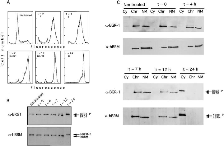
Association of BRG1 and hBRM with the nuclear matrix in synchronized HeLa cells. (A) HeLa Cells were synchronized by treatment with thymidine for 40 h. After release of the block (t = 0), samples were taken at the indicated time for flow cytometry analysis. 12 h later nocodazole was added at 0.1 μg/ml final concentration. (B) Synchronized cells were taken at the indicated time and total extracts were prepared. Equal quantities of protein were subjected to SDS-PAGE in 5% acrylamide gels and immunoblotted with α-BRG1 and α-hBRM antibodies. Phosphorylated and unphosphorylated BRG1 and hBRM are indicated. (C) Synchronized cells were taken at the indicated time and fractionated as described in Fig. 2. Cytoplasmic (Cy), chromatin (Chr), and nuclear matrix (NM) fractions were electrophoresed in 5% acrylamide gels and immunoblotted with α-BRG1 and α-hBRM antibodies.

