
Figure 8.
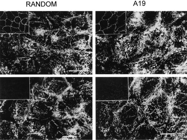
Effect of antisense A19 oligonucleotide on the apical distribution of apical tubulin in CACO-2 cells. The cells were continuously grown in random (a and c; control) or antisense A19 (b and d) oligonucleotides on glass coverslips for 9 d, saponin permeabilized, and fixed. The tight junction component ZO-1 (insets) and tubulin were localized by indirect immunofluorescence using specific second antibodies coupled to fluorescein and Texas red, respectively. Laser confocal optical sections were taken simultaneously in the green (ZO-1; insets) and red at two different levels: through the apical cytoplasm at the deep side of the ZO-1 signal (a, b) and ∼1 μm above the basal membrane (c, d). The pairs a–c and b–d correspond respectively to the same fields at different focal planes. Arrowheads point at tubulin clusters. Bars, 10 μm (equivalent to 30.4 μm for the insets).