
Figure 5.
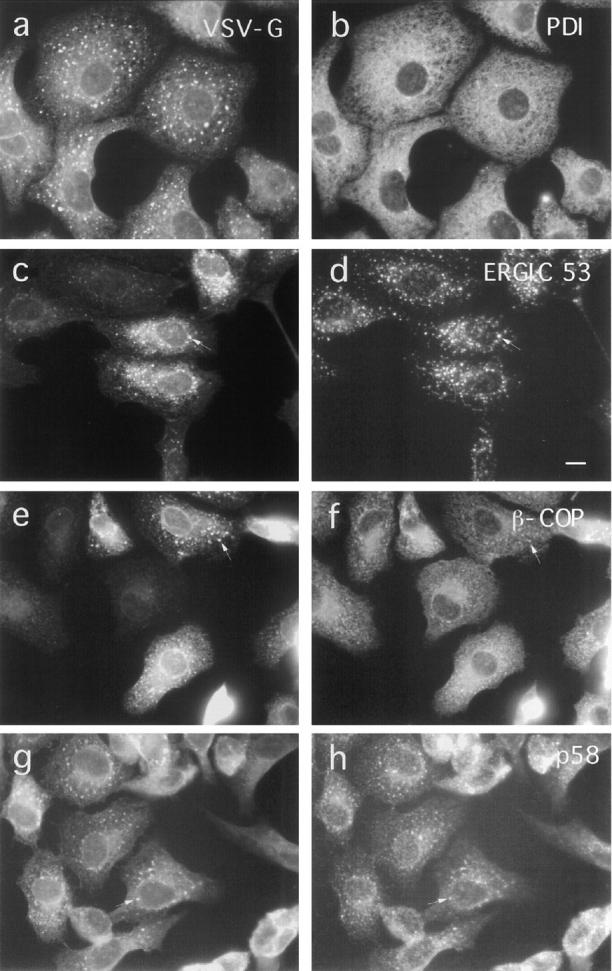
Effect of BFA on the distribution of ER, IC, and Golgi complex-associated proteins in VSV-infected cells. ts-O45 VSV-infected Vero cells were incubated with 5 μg/ml BFA for 2.5 h at 39.5°C and then fixed and double stained for ts-O45-G (a, c, e, and g) and the ER protein PDI (b), the IC marker protein ERGIC 53 (d), β-COP (f), and Golgi membrane associated p58 (h). ts-O45-G accumulates in patches enriched in ERGIC53, β−COP, and p58; PDI maintains its typical ER distribution and is not affected by BFA. Bar, 10 μm.