
Figure 2.
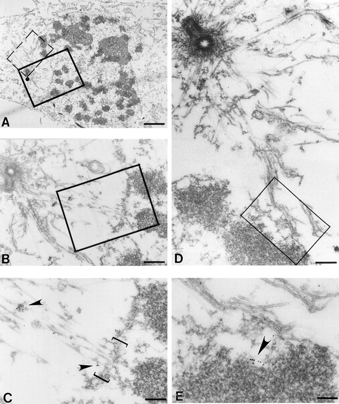
An associated plus end motor activity trafficks CENP-E along newly assembled astral microtubules into the nuclear domain after nuclear envelope fragmentation. HeLa cells were processed as described in Materials and Methods. (A) Low magnification view of a prophase/prometaphase HeLa cell bearing condensed chromosomes and a partially fragmented nuclear envelope. One spindle pole is readily apparent (asterisk). Examination of serial sections did not reveal another pole, consistent with a prophase cell before centriole separation. (B) Magnified view of boxed region in A shows that astral microtubules emanating from the centriole come in close proximity to the nuclear envelope. (C) Higher magnification view reveals that CENP-E is microtubule-associated along astral microtubules adjacent to the remaining nuclear envelope (bracket). Arrowheads point to microtubule-bound gold particles reporting CENP-E location. (D) Magnified view of the dashed box in A and highlighting astral microtubules passing through the fragmented lamina and lying in close proximity to a chromosome. (E) Higher magnification of the area boxed in D, revealing that some CENP-E is found along the microtubules, but additional CENP-E is associated with a localized domain on the chromosome, presumably the developing kinetochore. Note the chromosome is not yet attached to microtubules. Bars: (A) 2 μm; (B) 400 nm; (C) 200 nm; (D) 800 nm; (E) 140 nm.