
Figure 3.
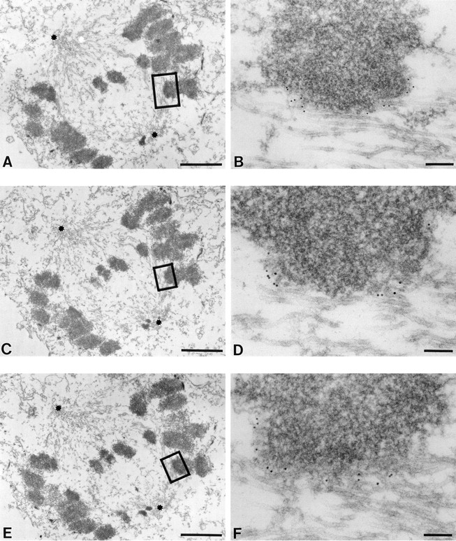
At early prometaphase, CENP-E binds all long the outermost surface of monooriented kinetochores attached laterally to spindle microtubules. HeLa cells grown on coverslips were preextracted and fixed. The visualization of CENP-E was achieved by 10-nm gold–conjugated goat anti–rabbit IgG. (A, C, and E) Low magnification serial sections of an early prometaphase HeLa cell. Asterisks denote the two poles of the developing bipolar spindle. An apparently monooriented chromosome is boxed, and higher power views are shown in B, D, and F. 10-nm gold particles representing CENP-E position decorate the interface between immature kinetochore and the laterally attached spindle microtubules. Note the labeling of CENP-E on the kinetochore appears as a crescent (C) shape. Bars: (A, C, and E) 2 μm; (B) 160 nm; (D and F) 110 nm.