
Figure 4.
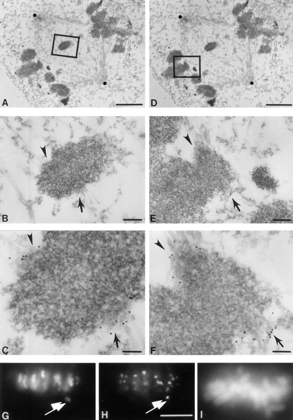
The leading kinetochore of a congressing chromosomes pair has increased level or accessibility of CENP-E. HeLa cells were processed as described in Fig. 2. (A and D) Low magnification views of a prometaphase HeLa cell (poles of the bipolar spindle are labeled with asterisks). (B and E) Intermediate magnification of two examples of a bioriented chromosome. (B) Boxed area of A showing chromosomes pair partially congressed from spindle poles toward the equator of spindle poles, but not yet aligned at the equator. (C and F) High magnification of the two bioriented chromosomes in B and E. 10-nm gold particles representing CENP-E decorate the outer kinetochore surface. A trilaminar structure of the kinetochore is not yet apparent, indicating that these kinetochores are not fully mature. (G–I) Double immunofluorescence demonstrating preferential CENP-E staining on the kinetochore closest to the midzone on a chromosome not yet congressed to the metaphase plate. (G) CENP-B, (H) CENP-E, and (I) DAPI to display chromosome positioning. Bars: (A and D) 2 μm; (B and E) 230 nm; (C) 90 nm; (F) 110 nm; (H–I) 10 μm.