
Figure 5.
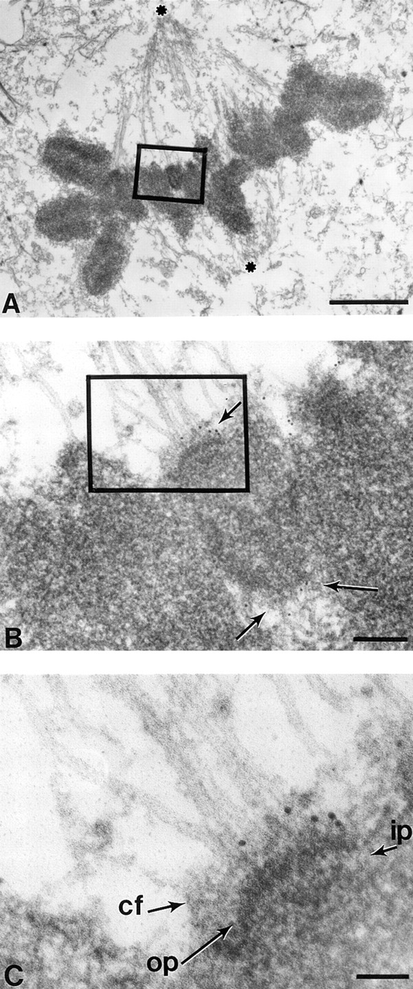
At metaphase CENP-E extends from the kinetochore outer plate at least 50 nm along spindle microtubules. Low magnification view of a metaphase HeLa cell with chromosomes aligned at the equator between the spindle poles (asterisks). (B) Magnified view of one metaphase chromosome showing that spindle microtubules indeed associate with a kinetochore with a trilaminar structure. Five 10-nm gold particles are located to each sister kinetochore (arrows). Five additional gold particles just to the right of the boxed area represent CENP-E associated with the kinetochore of another chromosome (more clearly seen in adjacent sections). (C) Higher magnification view shows that CENP-E is located to the corona fibers of the kinetochore. op, outer plate; ip, inner plate; cf, corona fibers. Bars: (A) 2 μm; (B) 170 nm; (C) 70 nm.