


Figure 7.
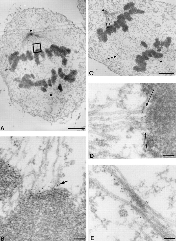
CENP-E remains a component of the kinetochore fibers as sister chromatids move toward the poles in anaphase. HeLa cells were processed as described in Fig. 2. (A) Low magnification view of an early anaphase HeLa cell. The two spindle pole positions are marked with asterisks (one is apparent while another is in a different section). (B) Magnified view of a kinetochore–microtubule interface shows that CENP-E is located between the kinetochore outer plate and the spindle microtubules (arrow). No gold particles are seen in other regions on chromosomes or on microtubules. (C) Low magnification view of a late anaphase HeLa cell bearing elongated spindle poles, labeled with asterisks; one is apparent while another is in a different section. Interzonal microtubules are readily seen (arrow). (D) Magnified view of the upper boxed region in C, showing that CENP-E is located between a kinetochore and its associated spindle microtubules (arrow). Examination of the number of particles revealed a reduction relative to metaphase. (E) Magnified view of area pointed by the arrow in C. Some CENP-E is now localized to the interzonal microtubules. Bars: (A) 2 μm; (B) 120 nm; (C) 2 μm; (D) 70 nm; (E) 90 nm.