
Figure 8.
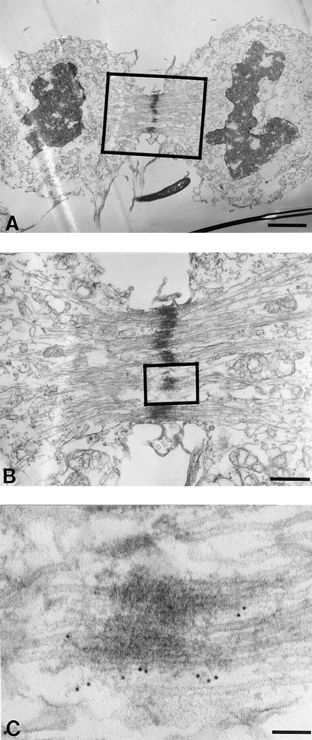
CENP-E cross-links the interzonal microtubules during telophase. HeLa cells were processed as described in Fig. 2. (A) Low magnification view of a late telophase HeLa cell. Lamin deposition to reform nuclei is partially complete. (B) Magnified view of boxed area in A shows that CENP-E is located along and/ or between the interzonal microtubules (boxed). (C) Higher magnification of interzonal microtubules shows that gold particles are primarily located between the microtubule bundles. Bars: (A) 2 μm; (B) 500 nm; (C) 90 nm.