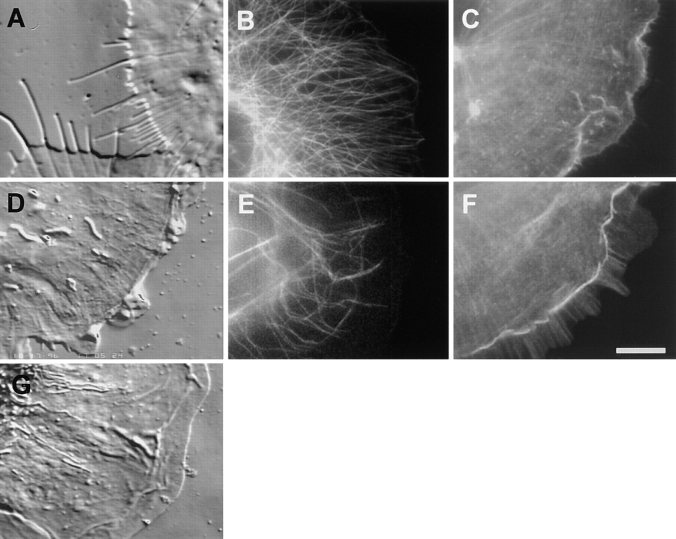
Figure 7.
Affects of cytochalasin D and BDM on the architecture and cytoskeleton of the lamella and lamellipodia. VE-DIC images of the lamella of living cells (A and D) ∼5 min after the perfusion of 2.5 μM cytochalasin D (A) or 20 mM BDM (D) and an untreated cell (G), for comparison. In A, the leading edge formed protrusions as the plasma membrane retracted around growing MTs. In D, the lamellipodia continued to ruffle and exhibit retrograde flow, but this motility ended abruptly at the margin at the base of the lamellipodia. Within the lamella, retrograde flow was inhibited. Fluorescence images of microtubules (B and E, with anti-tubulin antibodies) and F-actin (C and F, with Texas red-phalloidin) in fixed cells treated for 20 min with 2.5 μM cytochalasin D (B and C) or for 5 min with 20 mM BDM (C and F). Treatment with cytochalasin D caused the lamellipodia to fill up with microtubules and for F-actin to concentrate into large puncta. Preservation of the membrane extensions shown by VE-DIC in A by fixation was not possible. Treatment with BDM left the microtubule array slightly bundled but relatively undisturbed and resulted in a large concentration of F-actin at the actin marginal band. Bar, 10 μm.

