
Figure 4.
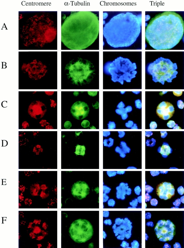
Centromere movement during polyploidizing megakaryocytes. TPO-treated primary megakaryocytes were stained with anticentromere antibody (red, first column), anti–α-tubulin antibody (green, second column), DAPI (blue, third column), and triple staining (fourth column) during mitosis. (A) Megakaryocyte in interphase. (B) Megakaryocytes in prometaphase. (C) Megakaryocyte with ploidy 8N at the stage just before metaphase. (D) Megakaryocyte with ploidy 8N in metaphase. (E) Megakaryocyte with ploidy 8N in anaphase A. (F) Megakaryocyte with ploidy 16N in anaphase A.