
Figure 8.
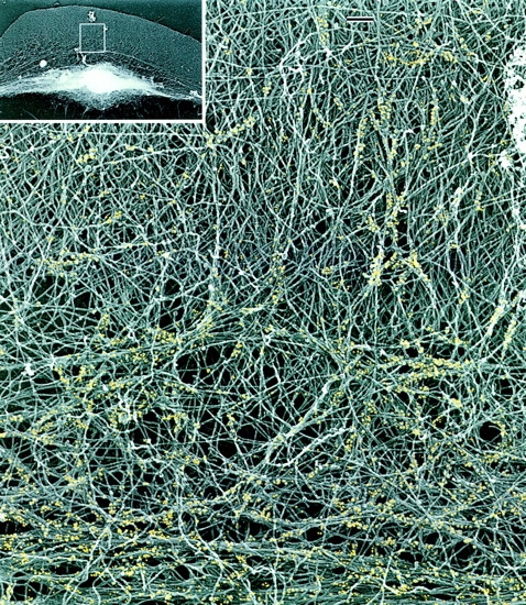
Relative distribution of actin and myosin II filaments in the keratocyte lamellipodia–cell body transition zone. EM of a detergent-extracted cell (overview in inset) after myosin immunogold labeling shows myosin filament clusters and the boundary bundle (bottom) within an actin filament network. Actin filaments forming small bundles and changing their course can be seen at sites of myosin localization. For better visualization, gold particles are digitally colorized in yellow. Bars, 0.2 μm.